Can I Get Social Security Disability Benefits for Back Pain and Spine Immobility?
- How Does the Social Security Administration Decide if I Qualify for Disability Benefits for Back Pain or Spine Impairments?
- About Back Pain and Disability
- Winning Social Security Disability Benefits for Back Problems by Meeting a Listing
- Residual Functional Capacity Assessment for Back Pain
- Getting Your Doctor’s Medical Opinion About What You Can Still Do
How Does the Social Security Administration Decide if I Qualify for Disability Benefits for Back Pain or Spine Impairments?
If you have a spine disorder that limits movement or causes chronic back pain, Social Security disability benefits may be available. To determine whether you are disabled by your back pain, or other spinal problems, the Social Security Administration first considers whether your back problems are severe enough to meet or equal a listing at Step 3 of the Sequential Evaluation Process. See Winning Social Security Disability Benefits for Back Pain by Meeting a Listing. If you meet or equal a listing because of back pain or other spine disorders, you are considered disabled. If your back problems are not severe enough to equal or meet a listing, Social Security Administration must assess your residual functional capacity (RFC) (the work you can still do, despite your back), to determine whether you qualify for benefits at Step 4 and Step 5 of the Sequential Evaluation Process. See Residual Functional Capacity Assessment for Back Pain and Spine Impairments.
About Back Pain and Disability
Impairments Causing Back Pain and Spine Immobility
Allegations of disability based on “back pain” are extremely common. Back pain and movement problems may be caused by a number of disorders including:
- Osteoarthritis (OA)
- Degenerative disc disease (DDD)
- Herniated nucleus pulposus (HNP) or herniated disc
- Osteoporosis
- Trauma
- Tumor
- Arachnoiditis
- Lumbar strain
- Spondylolisthesis
- Spinal stenosis
- Scoliosis
- Kyphosis
- Osteomyelitis
Some people may have structural problems in the spine that limit function (i.e., walking, bending, stooping, etc.). But question of disability usually depends on how much your chronic pain interferes with your ability to function (i.e., walk, bend, stoop, twist, lift, etc.). The great majority of individuals—more than 80%—who have acute low back pain from a strain of the ligaments and other soft-tissue supportive structures of the spine will recover within several months, even if they receive no treatment. Other individuals have a more chronic problem.
Spine Anatomy
The spine (vertebral column) has:
- 7 cervical (neck) vertebrae.
- 12 thoracic (chest, dorsal) vertebrae.
- 5 lumbar (lower back) vertebrae.
- 5 sacral vertebrae (fused triangular bone).
- 3 or 4 little vertebrae fused into a coccyx at the lower end of the spinal column.
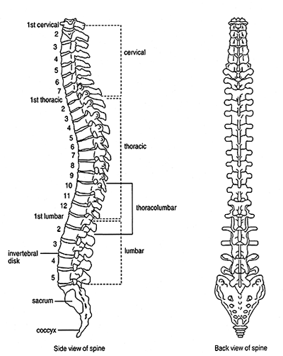
Figure 1: The human spine.
The spine provides structural support for the body and protects the spinal cord. Thirty-one pairs of nerve roots exit the spinal cord to form the peripheral nerves to the rest of the body. The peripheral nerves are sensory (carrying sensation), and motor (causing muscle movement). Disease processes affecting the spine can damage peripheral nerves at or near their origin (nerve roots), as well as the spinal cord itself.
Assessing Back Pain
The severity of back pain cannot be deduced solely based on abnormalities that are seen on plain X-rays, computerized tomography (CT), or magnetic resonance imaging (MRI) of the spine. Many people with significant degenerative abnormalities on X-ray have minimal or no symptoms, while some people who allege incapacitating back pain have minimal objective abnormalities. Nevertheless, even taking individual differences into account, there is a general correlation between objective abnormalities and credible pain.
The Social Security Administration will weigh your objective abnormalities, your reported pain and other symptoms, and your credibility in determining the severity of your impairment. In addition to objective evidence, your credibility with the Social Security Administration is strongly influenced by your behavior in seeking relief of alleged symptoms, your activities that are limited by pain, the nature and frequency of your visits to a doctor for treatment, your response to treatment given, and comments about your credibility in the treating doctor’s records.
Psychological and Social Factors in Back Pain
Although psychosocial factors play a major role in the functional loss caused by low back pain, there is no good way for the Social Security Administration to evaluate these factors. Psychosocial factors strongly predict future disability and the use of health care services for low back pain. Chronic disabling low back pain develops more frequently in patients who, at the initial evaluation for low back pain, have:
- A high level of “fear avoidance” (an exaggerated fear of pain leading to avoidance of beneficial activities);
- Psychological distress;
- Disputed compensation claims;
- Involvement in a tort-compensation system; or
- Job dissatisfaction.
These psychosocial factors are particularly prevalent in persons with low back pain for whom imaging shows only degenerative changes; 70 to 80 percent of such patients demonstrate psychological distress on psychometric testing or have disputed compensation issues, compared with 20 to 30 percent of patients whose imaging studies reveal definite pathologic or destructive processes. These psychosocial factors should be routinely assessed in patients with low back pain and taken into account in decisions regarding treatment.
Some degree of osteoarthritis of the spine is common in middle-aged people, even if they are not aware of it. OA of the spine can take several forms. In ankylosis, parts of the spine are abnormally fused together as a result of bony overgrowth. For example, bony spurs can fuse vertebral bodies together. The peripheral nerves formed from the spinal cord exit the bony spine through recesses in vertebrae called intervertebral foramina (see Figure 2 below). Some of these foramina can become encroached by osteoarthritis and require surgical decompression. Vertebrae have contact points with other vertebra called facet joints (see Figure 3 below).Arthritis affecting these facet joints can be painful and limit the motion of the spine. See also Can I Get Social Security Disability Benefits for Arthritis and Joint Damage?
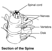
Figure 2: Spinal cord and nerve roots.
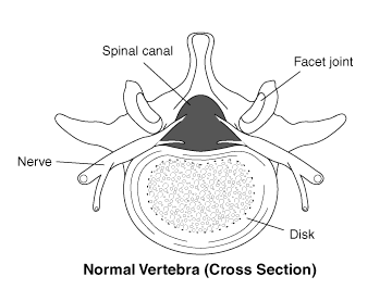
Figure 3: Normal spinal canal with facet joints.
Degenerative Disc Disease (DDD)
Degenerative disc disease refers to dehydration and shrinkage of the intervertebral discs that cushion the vertebral bodies of the spine. DDD is common and causes no symptoms in many older individuals. Everyone over about the age of 50 has some degree of DDD, which may or may not be symptomatic and functionally limiting. Osteoarthritis of the spine is frequently accompanied by DDD, while DDD without associated OA is also common. DDD can be seen on X-rays, MRI, and CT scans of the spine. It appears as narrowing of the space between vertebral bodies. Symptomatic DDD occurs between the 5th lumbar vertebra and the 1st sacral vertebra (L5-S1).
Sometimes a combination of OA and DDD produces enough symptoms that surgical fusion is performed in the lumbar spine (lumbar fusion) or cervical spine (neck). This procedure is done in an attempt to stabilize the spine and decrease pain. The surgery requires taking strips of bone from the posterior (back) upper part of the pelvic bone and laying them over the vertebral bodies that need to be stabilized (see Figure 4 below). Bone is living tissue and will incorporate the vertebral bodies into one solid mass. Sometimes, the bone strips do not incorporate well and the surgical fusion partially or wholly fails. Some fusions involve only two vertebrae, but multiple vertebrae may also be fused.
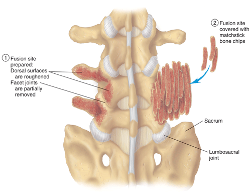
Figure 4: Vertebral fusion using bone strips.
Herniated Nucleus Pulposus (HNP) or Herniated Disc
A herniated nucleus pulposus is the protrusion of the hard, cartilaginous center (nucleus) of an intervertebral disk through the outer fibrous tissue (annulus fibrosa) (see Figures 5 and 6 below). Many small HNPs will produce acute symptoms that improve with time. Injection of corticosteroid drugs in the area of the HNP can also help relieve inflammation and pain. Some claimants have a large HNP that presses on a spinal nerve root, and must have part of the HNP removed (discectomy, diskectomy).
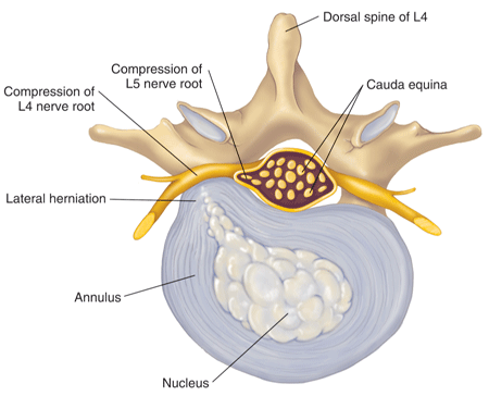
Figure 5: An intervertebral disk with lateral herniation.
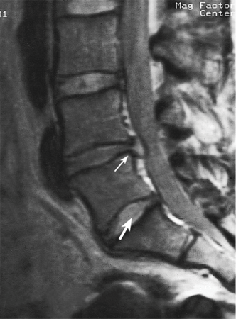
Figure 6: MRI view of herniated discs.
Often, a part of a vertebral body, the lamina, is also removed for surgical access and this procedure is known as a laminectomy. The problem with surgery around spinal nerve roots is that manipulation of tissues often leads to scarring that then again pressures the nerve root. This is particularly likely when a person has had multiple back surgeries. Many claimants who complain of chronic back pain and have a history of back surgery near nerve roots have scarring that can be identified on CT or MRI scans. In the absence of trauma, most HNPs occur in the lumbar (lower) spine, especially at the level of L5-S1.
Osteoporosis is a metabolic disorder associated with decrease in the mass of bone. By far, most of the instances of osteoporosis seen by the Social Security Administration are in post-menopausal women. Osteoporosis may be confined to the spine, but other bones may be involved in those who have used corticosteroids. A collapse of vertebral bodies, especially in the upper back, is known as a compression fracture. Compression fractures are visible on plain X-ray or other imaging studies. If the fracture involves the anterior (front) part of a vertebral body more than the rest of the vertebra, the spine will tend to curve forward and result in the popularly known dowager’s hump.
Compression fractures are graded in regard to the percent of the vertebra that is compressed, compared to the normal height of the vertebra. Normal height is judged from adjacent vertebrae. Pain, loss of motion, and muscle spasm are most likely to be present at the time of fracture and in the healing period. Marked or multiple compression fractures are more likely to produce chronic pain.
Plain X-rays are much less sensitive than bone densitometry in determining the severity of osteoporosis. A normal appearance of bone on plain X-rays only rules out the most advanced osteoporosis. Plain X-rays are fine for determining the percentage of vertebral body collapse in compression fractures.
Fractures of the bony spine are most commonly related to automobile or motorcycle accidents. There may be associated spinal cord injury. Traumatically fractured vertebrae are treated with a combination of surgical fusion and sometimes stabilization with metal rods.
The most serious tumors of the spine arise from cancer that has spread to the spine from breast, colon, prostate, or other origin. Tumors can not only cause chronic pain, but result in spinal fractures as they destroy bone. The spread of cancer of any kind to the spine is a serious development that must also be considered under the listings dealing with cancer.
Arachnoiditis is inflammation of some part of the arachnoid membrane that covers the spinal cord. It can produce severe chronic pain. Arachnoiditis may occur as a result of infection, but most commonly is seen after surgical procedures and use of contrast material to enhance visualization of structures with X-rays during myelography. Some people are more sensitive than others to contrast material. An MRI scan has about a 90% chance of showing this abnormality if it is present. A negative MRI scan for arachnoiditis is a strong argument that it is not present.
Lumbar strain refers to stress on the ligaments, muscles, and other soft tissues near the spine with resultant pain. There may or may not be underlying arthritis or DDD. Acute strain, associated with a particular lifting event, will almost always resolve in several months. If the pain is marked, there is associated muscle spasm and difficulty bending the back. When back pain continues for a prolonged period, orthopedists and other doctors tend to apply the diagnosis of “chronic lumbar strain,” if there is no other underlying identifiable abnormality that can be seen on imaging studies.
Spondylolisthesis is a slippage of vertebral bodies out of their normal position, usually a forward slippage of the 5th lumbar vertebra over the 1st sacral vertebra (L5-S1). More rarely, a type of spondylolisthesis called retrolisthesis involving the backward displacement of a vertebral body occurs. Most spondylolisthesis is seen in the lumbar spine (L1-L5/S1). This disorder can been seen on plain X-rays. It is significantly more likely to be seen on X-rays taken in the standing position than in those taken in lying position—with weight on the spine, slippage is more likely.
However, severe or even significant neurological abnormalities (sensory changes, reflex changes, muscle weakness or atrophy) are not to be expected in spondylolisthesis.
Studies have shown that most individuals with spondylolisthesis, lead active lives with little, if any, adjustment for having this type of spinal abnormality. Spondylolisthesis is most likely to become limiting as a contributing factor for spinal stenosis in combination with other spinal disorders, such as severe osteoarthritis and severely bulging intervertebral discs.
Spinal stenosis is a narrowing of the space inside the bony spine (see Figure 7 below), which sometimes results in pressure on the spinal cord and peripheral nerve roots from the spinal cord. The Social Security Administration most commonly sees such cases in claimants who have severe osteoarthritis of the lower spine. Spinal stenosis can be worsened by bulging or herniated disks (HNPs) (see Figure 8 below) and spondylolisthesis.
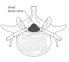 Figure 7: Spinal stenosis with a narrowing of the spinal canal.
.gif) Figure 8: Spinal stenosis caused by a herniated disk and osteoarthritis.
Figure 8: Spinal stenosis caused by a herniated disk and osteoarthritis.
Spinal stenosis most commonly involves the lower back, specifically the area somewhere between the 3rd lumbar vertebra and the beginning of the sacrum (L3-4, L4-5, and L5-S1 levels). Less frequently, the neck (cervical spine) may be involved with spinal stenosis; its presence in the upper back (thoracic spine) is rare.
Spinal stenosis is one of many possible causes of damage to the spinal cord (myelopathy). Myelopathy may be irreversible. Surgical decompression of the spinal cord may be necessary for severe cases, but even after surgery symptoms may not improve.
Pain and neurological abnormalities can be debilitating if treatment is not effective. Standing, walking, lifting, and carrying should be limited to weight that does not produce symptoms. A person with lumbar stenosis may have no symptoms during a physical examination, but may have severe symptoms with exertion.
Pain, weakness, numbness or other symptoms related to spinal stenosis usually appear gradually over a period of months or years. Symptoms are rapidly worsened by walking, lifting, jarring, carrying or other activities that strain the spinal structures. Sensory abnormalities, such as numbness, will occur before the onset of weakness. Symptoms are lessened or relieved by bending forward (including crouching) or lying. These symptoms are referred to as pseudoclaudication by the Social Security Administration, but are often also called neurogenic claudication.
In addition to osteoarthritis, causes of spinal stenosis include congenital spinal deformities (scoliosis, kyphosis, or congenital skeletal dysplasias like achondroplastic dwarfism); acquired deformities such as post-traumatic spinal fractures; inflammatory spinal diseases like ankylosing spondylitis; or stenosis may be of unknown cause. Tumors or infection present possible reversible causes of lumbar stenosis.
Spinal stenosis can be seen on imaging studies such as myelography, CT, and MRI scans. But myelography and CT scans can miss some types of stenosis.
Scoliosis is a sideways curvature to the spine (see Figure 9 below), associated with pain but not neurological impairment. Scoliosis can be of any degree of severity. Often the scoliosis is associated with one leg being shorter than the other. In these instances, the pelvis is not level causing the abnormal sideways spinal curvature. Scoliosis should be suspected with leg length discrepancies of 2.2 cm or greater.
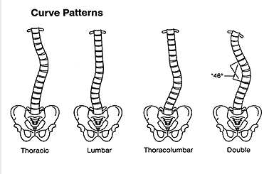
Figure 9: Varying degrees and types of scoliosis.
The abnormal spinal curve of scoliosis is measured on plain X-rays. The measurement is called a Cobb angle. Scoliosis may be considered present with abnormal curves greater than 10 degrees. Mild cases have angles less than 30 degrees.
Angles of 20 degrees or less are usually produce no symptoms. Bracing is prescribed for angles over 20 degrees. Curves over 40 degrees may produce neurological abnormalities such as sensory loss and weakness. Small curves of less than 30 degrees in childhood are not likely to get worse during adulthood, while more severe curves may continue to progress. Surgery with permanent rod implantation and fusion is indicated with curves greater than 45 degrees.
Claimants with curves 60 degrees or more require spirometry for restrictive lung disease (decreased vital capacity). See Can I Get Social Security Disability Benefits for Lung Disease? Heart failure may occur from scoliosis when abnormal curves are markedly severe at 100 degrees or more. Such cases are very rarely seen in Social Security disability adjudication.
During physical examination, a doctor may use a device known as a scoliometer (scoliosometer) to measure the degree of curvature. The scoliometer measurement should not be confused with the spinal curvature, although both are expressed in degrees. A scoliometer reading of 7 degrees corresponds to a 20-degree curve measured by Cobb angle on X-ray. Scoliometers are inexpensive and useful for screening, but direct curve measurements on plain X-ray views are needed for accurate determinations.
Kyphosis is an abnormal degree of curvature of the thoracic spine (upper back) in the forward direction (flexion). Kyphosis may be congenital or may occur in post-menopausal osteoporosis with collapse of the anterior (front) part of vertebral bodies in the upper back.
In kyphosis, forward curvature of the spine up to 20 degrees is considered normal, and mild up to 40 degrees. Bracing is prescribed for angles over 40 degrees and balance can be impaired by kyphotic curves greater than 40 degrees. Curves of 50 degrees or greater can produce a significant restrictive breathing deficit and should have vital capacity tested with spirometry. See Can I Get Social Security Disability Benefits for Lung Disease? As with scoliosis, extremely abnormal curves of 100 – 110 degrees or more can compromise cardiac function.
Osteomyelitis most often occurs as a result of trauma producing open wounds that allows the entry of bacteria into the body, as a result of surgical procedures, or as a result of bacteria circulating in the bloodstream—a condition known as bacteremia. Osteomyelitis of joints can affect their function by means of bone destruction and joint deformity. See Can I Get Social Security Disability Benefits for Arthritis or Joint Damage?
In weight-bearing bones, fractures through the area of infection can occur during the stage of acute infection, or later due to brittle bone. The orthopedic surgical management of osteomyelitis can be complex. Surgery may be needed to remove infected bone.
With modern antibiotics, acute osteomyelitis can be treated more effectively, so that chronic osteomyelitis is not as common as it was in the past. When chronic osteomyelitis does occur, it can present a difficult problem because the chronically infected bone may die and that restricts delivery of antibiotics through the bloodstream. Also, secondary infection may occur in tissues near the bone that involves different organisms than those that infect the bone itself.
An area of infected bone is called a sequestrum. In the treatment of chronic osteomyelitis, surgery to remove the sequestrum (sequestrectomy and curettage) along with infected soft tissues near the infection is a common requirement. Infected soft tissue removal may require reconstruction of soft tissues, such as muscle and skin grafts. The hole in the bone left by removal of the sequestrum may be packed with antibiotic beads. Antibiotic bead implantation may be temporary (10 days) to permanent, depending on the judgment of the surgeon. Whatever surgical antibiotic treatment is given, the patient will require prolonged systemic antibiotic therapy lasting well through surgical recovery, in order to prevent recurrent infection.
Infected bone fractures can be particularly difficult to heal. Such a situation might arise from an open wound and fractures occurring during an automobile accident or other trauma.
Continue to Winning Social Security Disability Benefits for Back Problems by Meeting a Listing.


